Ultrasonography
How do pregnancy ultrasounds work?
A probe is used to pass sound waves into the body. An image of the baby is
produced by the reflection of those sound waves. The images (sonogram)
are displayed on a television screen. The sonographer performing the
ultrasound can explain these images to you.Appropriate use of ultrasound poses no physical risk to you or your baby.
Why would you have a prenatal ultrasound?You may have an ultrasound to check the development of your pregnancy.
This includes:
- Confirming the stage of the pregnancy;
- Checking the continuation of a pregnancy if there has been bleeding;
Identifying a multiple pregnancy - Checking the position of the placenta
- Checking the amount of amniotic fluid
- Checking the physical development of the foetus
- Diagnosing birth defects and/or hereditary conditions.

A prenatal diagnostic ultrasound can detect physical abnormalities including:
- Congenital heart defects: Abnormalities of the structure and
function of the heart occur in 1 in 100 pregnancies - Limb reduction defects: Short or missing bones in the arm or leg
occur in 1 in 1000 pregnancies - Cleft lip palate: Failure of normal closure of tissue in the lip palate
area occurs in 1 in 1000 pregnancies - Neural tube defects: Abnormalities of the brain, skull, spine and
spinal cord
How is an ultrasound performed?
There are two ways in which obstetric ultrasound can be performed.
Abdominal ultrasound
- Abdominal ultrasound is the most common procedure and can be
used at any stage of pregnancy - A gel is applied to the abdomen to allow the sound waves to pass
from the transducer into the uterus - The bladder should be comfortably full to gain a clear ultrasound
picture.
Vaginal ultrasound
Vaginal ultrasound is less commonly performed. It is usually restricted to the first trimester (up to 12 weeks) of pregnancy. The bladder needs to be completely emptied in order to perform this method of ultrasound.
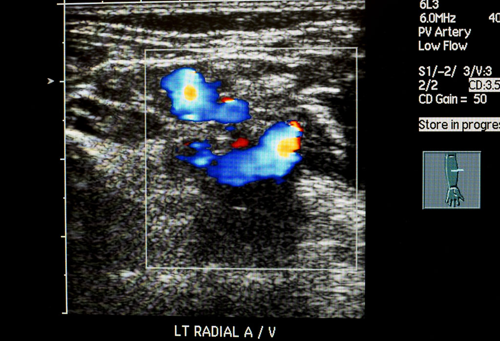
Color Doppler
Doppler Scan in Pregnancy
Doppler sonography is a technique which uses reflected sound waves to measure movements such as blood flow and heartbeat. Doppler ultrasound scans can be used to determine the speed of the blood flow and its direction. This information can be helpful in determining if the foetal growth is normal and whether the tissues are supplied with enough blood and nutrients.Doppler scans are performed with the same apparatus as a regular ultrasound scan and are normally used during the third trimester on women who have high-risk pregnancies.

What Is Doppler Scan?
A Doppler scan is similar to a regular ultrasound scan and works using high-frequency sound waves called ultrasound that aren’t audible to our ears. The ultrasound generated by the equipment bounces off bones and tissues like an echo and is recorded with a microphone. All of this is done with a small hand-held probe called a transducer. A gel which helps in the process is applied over the belly, and the transducer is pressed gently against the skin to scan. Denser substances like bones give off a better echo than the softer tissue that the body is made up of, and by comparing the echo, an image of the baby is generated in a computer and displayed in real time.What sets a Doppler scan apart is that unlike a regular ultrasound scan, it can detect the flow of blood in blood vessels, estimate the speed of the blood flow, determine its direction, detect blood clots, etc. Most ultrasound equipment these days has an inbuilt Doppler feature and both the scans can be done together.

Is It Safe To Have Doppler Scan?
Yes, like with all other ultrasound scans, Doppler is safe when done by trained professionals. The sonographer, who is the person carrying out the scans, follows a set of established guidelines that ensure that you and your baby are safe during the procedure. Since the scans work by using a focused beam of sound waves, the equipment can generate a small amount of heat as a consequence. Therefore, every ultrasound scan machine features a thermal index display that gives a rough estimation of how much heat is being generated.
The machines normally have a low thermal index and come with different output settings for different stages of pregnancy. Most of the ultrasound scans don’t exceed 30 minutes, and a typical Doppler scan lasts only a few minutes. This poses no risk to the baby or the mother. In decades of using ultrasound scans during pregnancies, there has been no evidence that suggests that these scans are harmful.
Why Is Doppler Scan Done During Pregnancy
Generally, women need two basic ultrasound scans during their pregnancy. The first one is during the first trimester to look for the number of babies, check for the baby’s heartbeat, to determine the baby’s growth and predict a due date. The second scan is done in the second trimester to check for physical abnormalities and confirm that the baby is developing normally.
If the doctor finds any anomaly during these scans, a Doppler scan is done for further investigation. Dopplers are often used to check the placental blood flow, the foetal umbilical blood flow, and blood flow in the heart and brain to ensure it is normal. If any restriction to the flow is detected, the doctors can determine if it is caused by a restricted blood vessel, sickle cell anaemia or RH sensitisation.Restricted blood flow in the foetus can cause lower birth weight, impaired development, reduced size, etc. A special type of Doppler ultrasound called a Transcranial Doppler is used to evaluate the risk of stroke in babies with sickle cell anaemia. Doppler scans are also recommended in conditions such as:
- Carrying twins or more
- The mother has a low or high body mass index (BMI)
- Medical conditions such as diabetes and high blood pressure
- The baby is affected by rhesus antibodies
- The baby’s growth rate is low
- Previous miscarriage or a small baby
- The mother smokes
When Will Your Doctor Ask For A Doppler Test
Doctors ask for Doppler test while pregnant when complications or abnormalities are detected in earlier scans that demand extra care for the pregnant woman during the course of pregnancy. Some of the other common conditions when doctors ask for Doppler scans are:
- Multiple pregnancies:
When the mother is carrying multiples, the pregnancy is considered a high risk one and is monitored regularly with Doppler scans. This is because such pregnancies have chances for several complications to arise. Some of them include – TTTS (Twin to Twin Transfusion syndrome), IUGR (Intra Uterine Growth Retardation), umbilical cord entanglement, etc. These complications can be detected early with a Doppler scan.
- Placental problems:
The placenta supplies the foetus with blood, nutrients and oxygen from the mother’s body. Healthy blood flow in the placenta is essential for normal development of the baby. During the second trimester’s anomaly detection scan, if problems such as slower foetal development are seen, a Doppler scan is used to detect any irregularities in the placental blood flow. A Doppler scan is also used if placenta previa (low lying placenta) is detected. The scan can show the placental position which can change toward the end of the pregnancy.
Health conditions of the mother:
The mother’s health has a profound effect on the growth of the foetus. Doctors use Doppler scans to determine the blood flow rate in the umbilical arteries and the placenta. There are conditions under which blood flow in the arteries can get restricted, such as the contraction of arteries due to smoking, certain medications, and other lifestyle related causes. The contracted arteries offer a high resistance to the flow of blood, resulting in improper oxygen and nutrient supply to the foetus. Conditions such as high blood pressure and diabetes can also have a significant effect on this.
- Health conditions of the foetus:
When the growth rate of the foetus in previous ultrasound scans is not satisfactory, doctors use a Doppler foetus scan for further analysis.
How Doppler Scan Is Different From Other Pregnancy Scans
A typical ultrasound scan bounces high frequency sound off the tissues of the body to give a thin 2 dimensional image which does not show any movement. A Doppler ultrasound scan on the other hand relies on the Doppler Effect. The phenomenon is a continuous change in the frequency of the bounced sound wave when it hits something that moves, like the blood flow in the arteries. It can measure the difference between blood that is moving away from the probe and blood that is moving towards the probe and also its speed. A Doppler can also detect the heartbeat of the foetus which other scans cannot.
